Contents Yves Clermont, Michael Lalli, Zsuzsanna Bencsath-Makkai
LIGHT-MICROSCOPIC HISTOLOGY ATLAS
     15. Male Reproductive System
  1     2     3     4     5     6     7     8     9     10     11     12     13     14     15     16     17     18     19     20     21     22     23     24     25  
  26     27     28     29     30     31     32     33     34     35     36     37     38     39     40     41     42     43     44  

    15.11   Sertoli cell Version française
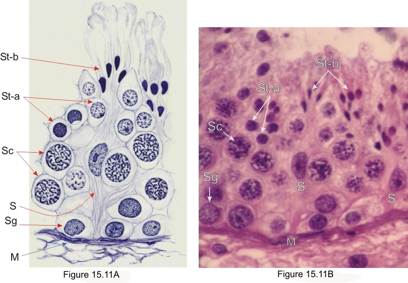
Human seminiferous epithelium containing a Sertoli cell (S) surrounded by germinal cells.

The drawing on the left produced by M. Oeltzschner is based on the image on the right.

The Sertoli cells are closely attached to the basement membrane (M) of the tubule, but their lateral cytoplasmic limits are not visible with the light microscope.

The germinal cells are labelled as follows:

  • Spermatogonia (Sg)
  • Spermatocytes (Sc)
  • Early round spermatids (St-a)
  • Late elongated spermatids (St-b)

Stain: H–­­E
Magnification: ×900


We'd very much appreciate receiving your comments and suggestions!We'd very much appreciate receiving your comments and suggestions!

Creative Commons Licence The text and images of this Histology Atlas, by Yves Clermont, Michael Lalli & Zsuzsanna Bencsath-Makkai, are licensed under a Creative Commons Attribution-Noncommercial-No Derivative Works 2.5 Canada Licence and cannot be modified without the written permission of the authors. Use of any text or images must carry an acknowledgement which includes a link to the original work.


HAweb/Zs B-M -> HAbase-generated on 2013-09-13