|
||
| 15. Male Reproductive System | ||
| 1 2 3 4 5 6 7 8 9 10 11 12 13 14 15 16 17 18 19 20 21 22 23 24 25 | ||
| 26 27 28 29 30 31 32 33 34 35 36 37 38 39 40 41 42 43 44 |
| |||
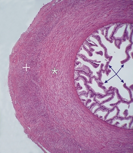 |
Transverse section of the ampulla of the vas deferens from a dog.
This ampulla is a dilatation of the vas deferens as it approaches the seminal vesicle and the prostate (see Figure 15.1). The large lumen of this duct is delimited by a thick mucosa showing numerous branching and interconnected folds (arrows). These folds are lined with a simple columnar epithelium. This mucosa is similar to that of the seminal vesicle shown below (see figures 33 to 37). The wall of the duct shows a thick muscular coat in which the smooth muscle fibres, which run circularly or longitudinally, form two distinct layers, the inner circular (*) and the outer longitudinal (+) layers. Stain: HE
|
||