Contents Yves Clermont, Michael Lalli, Zsuzsanna Bencsath-Makkai
LIGHT-MICROSCOPIC HISTOLOGY ATLAS
     15. Male Reproductive System
  1     2     3     4     5     6     7     8     9     10     11     12     13     14     15     16     17     18     19     20     21     22     23     24     25  
  26     27     28     29     30     31     32     33     34     35     36     37     38     39     40     41     42     43     44  

    15.35   Seminal vesicle Version française
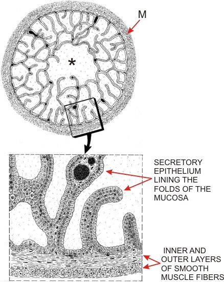
The top drawing shows a cross section of the simple tubular gland that composes a seminal vesicle.

Delimited by a tunica muscularis (M), the mucosa shows numerous branching and anastomotic folds forming spaces that connect with each other and with a central lumen (*). The details of the framed area are shown in the drawing Figure 15.36.

The folds of the mucosa have a core of lamina propria, made up of a well-vascularized connective tissue. They are lined with a simple columnar epithelium composed mainly of secretory cells and of a few basal cells.

These two drawings reproduce the two histological fields shown in Figure 15.36 and Figure 15.37.


We'd very much appreciate receiving your comments and suggestions!We'd very much appreciate receiving your comments and suggestions!

Creative Commons Licence The text and images of this Histology Atlas, by Yves Clermont, Michael Lalli & Zsuzsanna Bencsath-Makkai, are licensed under a Creative Commons Attribution-Noncommercial-No Derivative Works 2.5 Canada Licence and cannot be modified without the written permission of the authors. Use of any text or images must carry an acknowledgement which includes a link to the original work.


HAweb/Zs B-M -> HAbase-generated on 2013-09-13