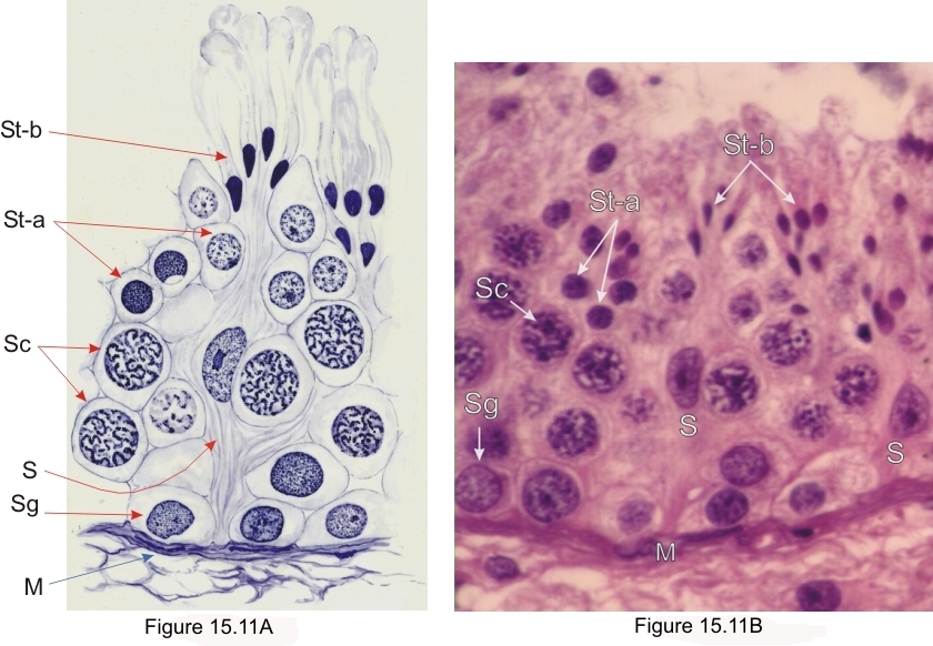
Sommaire Yves Clermont, Michael Lalli, Zsuzsanna Bencsath-Makkai
ATLAS D'HISTOLOGIE EN MICROSCOPIE OPTIQUE
     15. Appareil Reproducteur Mâle
  1     2     3     4     5     6     7     8     9     10     11     12     13     14     15     16     17     18     19     20     21     22     23     24     25  
  26     27     28     29     30     31     32     33     34     35     36     37     38     39     40     41     42     43     44  

    15.11   La cellule de Sertoli English version
L’épithélium séminifère humain montrant deux noyaux de cellules de Sertoli (S) entourés de cellules germinales.

La photo de droite a servi de modèle pour l’exécution du dessin de gauche réalisé par M. Oeltzchner.

Les cellules de Sertoli sont étroitement attachées à la membrane limitante du tube (M) mais leur membrane latérale n’est pas identifiable en microscope optique. Les cellules germinales sont étiquetées comme suit:

  • Les spermatogonies (Sg)
  • Les spermatocytes (Sc)
  • Les jeunes permatides rondes (St-a)
  • Les spermatides en fin de la spermiogenèse (St-b) aux noyaux condensés

Coloration: H–­­É
Grossissement: ×900


We'd very much appreciate receiving your comments and suggestions!We'd very much appreciate receiving your comments and suggestions!

Creative Commons Licence The text and images of this Histology Atlas, by Yves Clermont, Michael Lalli & Zsuzsanna Bencsath-Makkai, are licensed under a Creative Commons Attribution-Noncommercial-No Derivative Works 2.5 Canada Licence and cannot be modified without the written permission of the authors. Use of any text or images must carry an acknowledgement which includes a link to the original work.


HAweb/Zs B-M -> HAbase-generated on 2013-09-13