 |
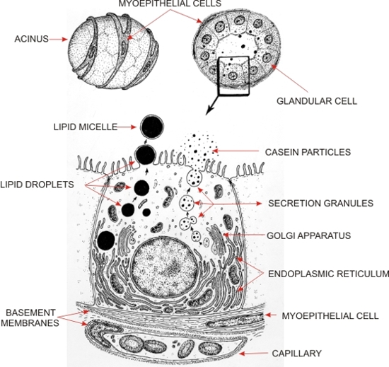
Drawing showing the acini of a lactating mammary gland in three dimensions (top left) to illustrate the myoepithelial cells at their surface and in transverse section (top right).
The framed epithelial cell with the underlying myoepithelial cell is magnified to show the organelles of the secretory cells as seen with the electron microscope.
This drawing illustrates the mode of formation of the two main types of secretions present in milk: lipid droplets (left) and casein particles (right). The lipid droplets are not limited by a membrane in the cytoplasm and are extruded at the apex of the cell surrounded by an envelope of apical cytoplasmic membrane. The casein particles accumulate in the cisternae of the Golgi apparatus and then collect in secretory granules. These granules migrate toward the apical membrane, with which they fuse, and liberate, by exocytosis, their particles of casein into the lumen of the acinus. Other substances such as proteins (e.g., lactalbumin) and sugars (e.g., lactose), included in secretory granules, are also released into the lumen of the acinus.
|