Contents Yves Clermont, Michael Lalli, Zsuzsanna Bencsath-Makkai
LIGHT-MICROSCOPIC HISTOLOGY ATLAS
     16. Female Reproductive system
  1     2     3     4     5     6     7     8     9     10     11     12     13     14     15     16     17     18     19     20     21     22     23     24     25  
  26     27     28     29     30     31     32     33     34     35     36     37     38     39     40     41     42     43     44     45     46     47  

    16.7   Primary follicle Version française
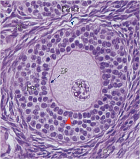
Primary multilaminar follicle (Pa-m) in the ovarian cortex.

The enlarged oocyte in the centre of the follicle shows the nucleus (N) in an abundant and granulated cytoplasm. This nucleus (N) contains diffuse chromosomes and a large nucleolus (*).

This oocyte is surrounded by several layers of follicular cells (Fo). Next to the oocyte, a layer of follicular cells (arrowhead) is closely applied to a thick eosinophilic layer. This layer, rich in glycoproteins, corresponds to the zona pellucida (ZP), which is deposited on the plasma membrane of the oocyte.

Numerous fibrocytes (Fi) of the stroma surround this growing follicle.

Stain: H–­­E
Magnification: ×1000


We'd very much appreciate receiving your comments and suggestions!We'd very much appreciate receiving your comments and suggestions!

Creative Commons Licence The text and images of this Histology Atlas, by Yves Clermont, Michael Lalli & Zsuzsanna Bencsath-Makkai, are licensed under a Creative Commons Attribution-Noncommercial-No Derivative Works 2.5 Canada Licence and cannot be modified without the written permission of the authors. Use of any text or images must carry an acknowledgement which includes a link to the original work.


HAweb/Zs B-M -> HAbase-generated on 2013-09-13