Contents Yves Clermont, Michael Lalli, Zsuzsanna Bencsath-Makkai
LIGHT-MICROSCOPIC HISTOLOGY ATLAS
     16. Female Reproductive system
  1     2     3     4     5     6     7     8     9     10     11     12     13     14     15     16     17     18     19     20     21     22     23     24     25  
  26     27     28     29     30     31     32     33     34     35     36     37     38     39     40     41     42     43     44     45     46     47  

    16.6   Ovarian cortex Version française
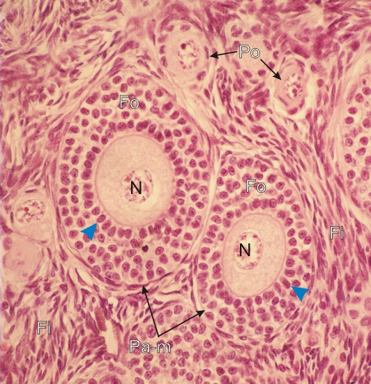
High-power view of the ovarian cortex.

This field shows a few primordial follicles (Po) and two primary multilaminar follicles (Pa-m).

These growing primary follicles show enlarging oocytes with a large nucleus (N). The oocytes are surrounded by several layers of small follicular cells (Fo). A distinct layer of follicular cells (arrowheads) is applied to the surface of the oocytes.

Numerous fibrocytes (Fi) in the stroma surround the various follicles.

Stain: H–­­E
Magnification: ×1000


We'd very much appreciate receiving your comments and suggestions!We'd very much appreciate receiving your comments and suggestions!

Creative Commons Licence The text and images of this Histology Atlas, by Yves Clermont, Michael Lalli & Zsuzsanna Bencsath-Makkai, are licensed under a Creative Commons Attribution-Noncommercial-No Derivative Works 2.5 Canada Licence and cannot be modified without the written permission of the authors. Use of any text or images must carry an acknowledgement which includes a link to the original work.


HAweb/Zs B-M -> HAbase-generated on 2013-09-13