Contents Yves Clermont, Michael Lalli, Zsuzsanna Bencsath-Makkai
LIGHT-MICROSCOPIC HISTOLOGY ATLAS
     16. Female Reproductive system
  1     2     3     4     5     6     7     8     9     10     11     12     13     14     15     16     17     18     19     20     21     22     23     24     25  
  26     27     28     29     30     31     32     33     34     35     36     37     38     39     40     41     42     43     44     45     46     47  

    16.12   Follicular atresia Version française
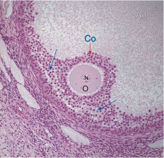
Section of a small portion of a large secondary or antral follicle (An) showing early signs of follicular degeneration or atresia.

The oocyte (O), with its centrally located nucleus (N), appears normal and is surrounded by a cumulus oophorus (CO) seemingly composed of normal-looking follicular cells.

Along the wall of the follicle, however, many follicular cells show signs of degeneration and death (arrows) characteristic of early follicular atresia.

Stain: H–­­E
Magnification: ×400


We'd very much appreciate receiving your comments and suggestions!We'd very much appreciate receiving your comments and suggestions!

Creative Commons Licence The text and images of this Histology Atlas, by Yves Clermont, Michael Lalli & Zsuzsanna Bencsath-Makkai, are licensed under a Creative Commons Attribution-Noncommercial-No Derivative Works 2.5 Canada Licence and cannot be modified without the written permission of the authors. Use of any text or images must carry an acknowledgement which includes a link to the original work.


HAweb/Zs B-M -> HAbase-generated on 2013-09-13