Contents Yves Clermont, Michael Lalli, Zsuzsanna Bencsath-Makkai
LIGHT-MICROSCOPIC HISTOLOGY ATLAS
     16. Female Reproductive system
  1     2     3     4     5     6     7     8     9     10     11     12     13     14     15     16     17     18     19     20     21     22     23     24     25  
  26     27     28     29     30     31     32     33     34     35     36     37     38     39     40     41     42     43     44     45     46     47  

    16.19   Corpus luteum Version française
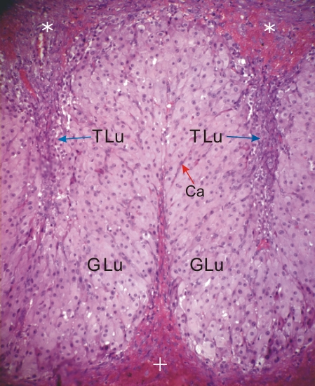
Section of a human corpus luteum.

The stroma (*) surrounding the ovary is at the top and the blood clot (+) in a central cavity of the corpus luteum is at the bottom.

The lightly stained steroid-secreting granulosa lutein cells (GLu) constitute the major cellular component of the organ.

At this magnification the smaller theca lutein cells (TLu) are difficult to identify but are present in the crevices between the granulosa lutein cells (GLu).

Collapsed capillaries (Ca) are indicated.

Stain: H–­­E
Magnification: ×300


We'd very much appreciate receiving your comments and suggestions!We'd very much appreciate receiving your comments and suggestions!

Creative Commons Licence The text and images of this Histology Atlas, by Yves Clermont, Michael Lalli & Zsuzsanna Bencsath-Makkai, are licensed under a Creative Commons Attribution-Noncommercial-No Derivative Works 2.5 Canada Licence and cannot be modified without the written permission of the authors. Use of any text or images must carry an acknowledgement which includes a link to the original work.


HAweb/Zs B-M -> HAbase-generated on 2013-09-13