Contents Yves Clermont, Michael Lalli, Zsuzsanna Bencsath-Makkai
LIGHT-MICROSCOPIC HISTOLOGY ATLAS
     16. Female Reproductive system
  1     2     3     4     5     6     7     8     9     10     11     12     13     14     15     16     17     18     19     20     21     22     23     24     25  
  26     27     28     29     30     31     32     33     34     35     36     37     38     39     40     41     42     43     44     45     46     47  

    16.8   Ovarian cortex Version française
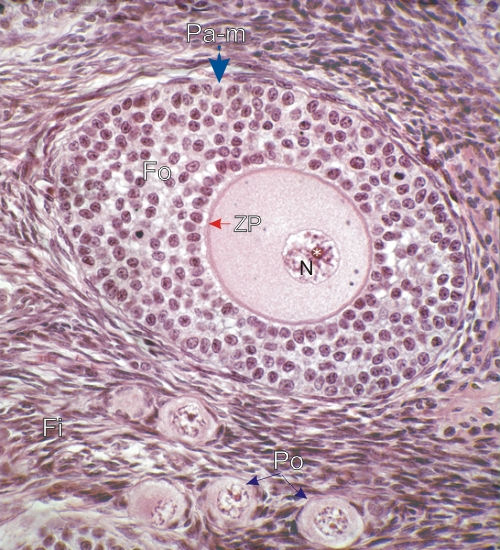
Ovarian cortex showing, next to a primary multilaminar follicle (Pa-m), several primordial follicles (Po).

This field permits a comparison of the marked difference in sizes of these two types of follicles.

Note the numerous follicular cells (Fo) of the primary follicle and the large size of the oocyte with its large nucleus (N) and nucleolus (*). The abundant cytoplasm of the oocyte is delimited by the zona pellucida (ZP).

The small primordial follicles (Po) show small oocytes with barely recognizable flattened follicular cells at their surfaces.

The fibrocytes (Fi) of the ovarian stroma are labelled.

Stain: H–­­E
Magnification: ×600


We'd very much appreciate receiving your comments and suggestions!We'd very much appreciate receiving your comments and suggestions!

Creative Commons Licence The text and images of this Histology Atlas, by Yves Clermont, Michael Lalli & Zsuzsanna Bencsath-Makkai, are licensed under a Creative Commons Attribution-Noncommercial-No Derivative Works 2.5 Canada Licence and cannot be modified without the written permission of the authors. Use of any text or images must carry an acknowledgement which includes a link to the original work.


HAweb/Zs B-M -> HAbase-generated on 2013-09-13