Contents Yves Clermont, Michael Lalli, Zsuzsanna Bencsath-Makkai
LIGHT-MICROSCOPIC HISTOLOGY ATLAS
     16. Female Reproductive system
  1     2     3     4     5     6     7     8     9     10     11     12     13     14     15     16     17     18     19     20     21     22     23     24     25  
  26     27     28     29     30     31     32     33     34     35     36     37     38     39     40     41     42     43     44     45     46     47  

    16.3   Ovary Version française
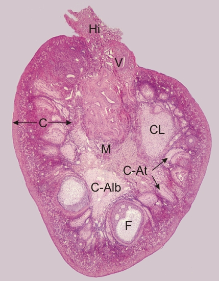
Transverse section of the ovary of a monkey.

This field shows large vessels (V) in the hilus (Hi) and medulla (M). These vessels enter and leave the ovary at the hilus.

In the cortex (C), the following structures can be identified:

  • A large growing ovarian follicle (F)
  • Several small connective tissue scars corresponding to residues of atretic follicles or corpora atretica (C-At)
  • A large corpus albicans (C-Alb) arising from a regressed corpus luteum (CL)

The other structures of the ovarian cortex are seen at higher magnifications in the subsequent figures.

Stain: H–­­E
Magnification: ×40


We'd very much appreciate receiving your comments and suggestions!We'd very much appreciate receiving your comments and suggestions!

Creative Commons Licence The text and images of this Histology Atlas, by Yves Clermont, Michael Lalli & Zsuzsanna Bencsath-Makkai, are licensed under a Creative Commons Attribution-Noncommercial-No Derivative Works 2.5 Canada Licence and cannot be modified without the written permission of the authors. Use of any text or images must carry an acknowledgement which includes a link to the original work.


HAweb/Zs B-M -> HAbase-generated on 2013-09-13