|
||
| 16. Female Reproductive system | ||
| 1 2 3 4 5 6 7 8 9 10 11 12 13 14 15 16 17 18 19 20 21 22 23 24 25 | ||
| 26 27 28 29 30 31 32 33 34 35 36 37 38 39 40 41 42 43 44 45 46 47 |
| |||
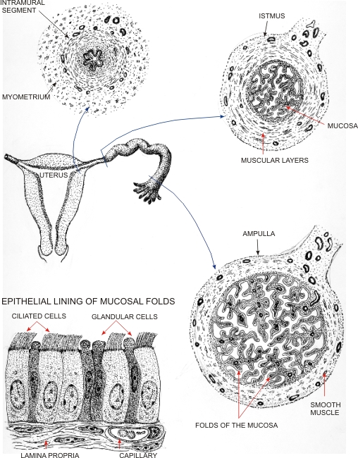 |
Drawings showing the main histological features of the oviduct or Fallopian tube.
Three cross sections of the oviduct are represented, i.e., the intramural or interstitial segment located in the wall of the uterus, the isthmus and the ampulla. Histological images of the mucosa of these three portions of the oviduct are shown in figures 16.22 to 16.28. The two main types of epithelial cells lining the mucosa of the ampulla are illustrated (bottom left). These are the glandular non-ciliated columnar cells and the ciliated columnar cells. While the apices of the ciliated cells show long motile cilia, the glandular cells do not show cilia but a cytoplasmic process covered with short microvilli (not shown).
|
||