Contents Yves Clermont, Michael Lalli, Zsuzsanna Bencsath-Makkai
LIGHT-MICROSCOPIC HISTOLOGY ATLAS
     16. Female Reproductive system
  1     2     3     4     5     6     7     8     9     10     11     12     13     14     15     16     17     18     19     20     21     22     23     24     25  
  26     27     28     29     30     31     32     33     34     35     36     37     38     39     40     41     42     43     44     45     46     47  

    16.16   Corpus atreticum Version française
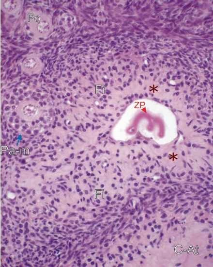
Section of a corpus atreticum of a monkey at an advanced stage of formation.

In a small central residual cavity of a regressed follicle, the collapsed, persistent, wavy zona pellucida (ZP) facilitates the identification of this corpus atreticum.

Surrounding this small cavity, many fibrocytes (Fi) are widely separated by lightly stained collagen (*). This image shows an early stage in the formation of a typical scar of a corpus atreticum (C-At) (bottom right), already seen in Figure 16.11.

A few primordial follicles (Po) and a primary multilaminar follicle (Pa-m) are seen in the surrounding ovarian stroma.

Stain: H–­­E
Magnification: ×500


We'd very much appreciate receiving your comments and suggestions!We'd very much appreciate receiving your comments and suggestions!

Creative Commons Licence The text and images of this Histology Atlas, by Yves Clermont, Michael Lalli & Zsuzsanna Bencsath-Makkai, are licensed under a Creative Commons Attribution-Noncommercial-No Derivative Works 2.5 Canada Licence and cannot be modified without the written permission of the authors. Use of any text or images must carry an acknowledgement which includes a link to the original work.


HAweb/Zs B-M -> HAbase-generated on 2013-09-13