Contents Yves Clermont, Michael Lalli, Zsuzsanna Bencsath-Makkai
LIGHT-MICROSCOPIC HISTOLOGY ATLAS
     5. Vessels
  1     2     3     4     5     6     7     8     9     10     11     12     13     14     15     16     17     18     19     20     21     22     23     24     25  
  26     27     28     29     30     31     32     33     34     35  

    5.34   Lymphatic vessel Version française
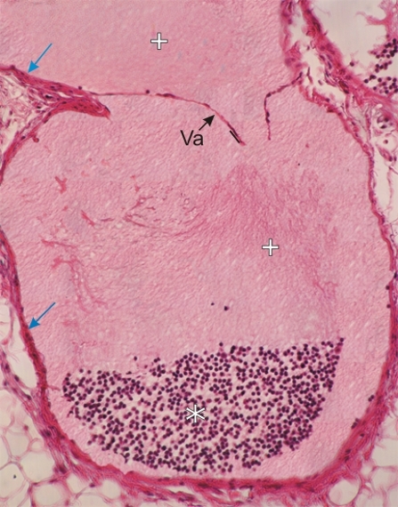
Section of a large lymphatic vessel from the thoracic cavity.

This field shows a longitudinal section of the vessel going through a valve (Va). This valve is composed of two leaflets of endothelium sitting on a thin layer of connective tissue. The two leaflets of the valve are facing spaces (+) containing lymph. In the lower part of the field there is an accumulation of lymphocytes (*).

The wall of the lymphatic mainly composed of connective tissue is indicated (arrows)

Stain: H–­­E
Magnification: ×200


We'd very much appreciate receiving your comments and suggestions!We'd very much appreciate receiving your comments and suggestions!

Creative Commons Licence The text and images of this Histology Atlas, by Yves Clermont, Michael Lalli & Zsuzsanna Bencsath-Makkai, are licensed under a Creative Commons Attribution-Noncommercial-No Derivative Works 2.5 Canada Licence and cannot be modified without the written permission of the authors. Use of any text or images must carry an acknowledgement which includes a link to the original work.


HAweb/Zs B-M -> HAbase-generated on 2013-09-13