|
||
| 5. Vessels | ||
| 1 2 3 4 5 6 7 8 9 10 11 12 13 14 15 16 17 18 19 20 21 22 23 24 25 | ||
| 26 27 28 29 30 31 32 33 34 35 |
| |||
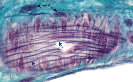 |
Tangential section of an arteriole stained with Massons trichrome and iron hematoxylin to demonstrate elastin.
The plane of section goes through the internal elastic layer and shows a face view of elastic fibres running longitudinally (blue arrows). The underlying smooth muscle cells are seen side by side and arranged circularly in the media. Around the media, the dense connective tissue of the adventitia (*), stained green, is composed of type I collagen and of a few elastic fibres. The nuclei of fibrocytes (F) in the adventitia are indicated. Stain: Massons Trichrome
|
||