Contents Yves Clermont, Michael Lalli, Zsuzsanna Bencsath-Makkai
LIGHT-MICROSCOPIC HISTOLOGY ATLAS
     5. Vessels
  1     2     3     4     5     6     7     8     9     10     11     12     13     14     15     16     17     18     19     20     21     22     23     24     25  
  26     27     28     29     30     31     32     33     34     35  

    5.33   Lymphatic vessels Version française
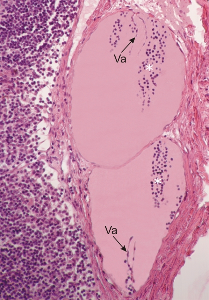
This section shows two lymphatic vessels within the connective tissue capsule of a lymph node.

These vessels contain small lymphocytes (*) and lymph free of red blood cells. They are lined with an endothelium sitting on a thin layer of connective tissue.

In both lymphatics there are membranous projections of their wall that form small valves or valvules (Va). These valves favour the anterograde flow of lymph along the lymphatic vessels

Stain: H–­­E
Magnification: ×200


We'd very much appreciate receiving your comments and suggestions!We'd very much appreciate receiving your comments and suggestions!

Creative Commons Licence The text and images of this Histology Atlas, by Yves Clermont, Michael Lalli & Zsuzsanna Bencsath-Makkai, are licensed under a Creative Commons Attribution-Noncommercial-No Derivative Works 2.5 Canada Licence and cannot be modified without the written permission of the authors. Use of any text or images must carry an acknowledgement which includes a link to the original work.


HAweb/Zs B-M -> HAbase-generated on 2013-09-13