Contents Yves Clermont, Michael Lalli, Zsuzsanna Bencsath-Makkai
LIGHT-MICROSCOPIC HISTOLOGY ATLAS
     5. Vessels
  1     2     3     4     5     6     7     8     9     10     11     12     13     14     15     16     17     18     19     20     21     22     23     24     25  
  26     27     28     29     30     31     32     33     34     35  

    5.23   Arteriole and venule Version française
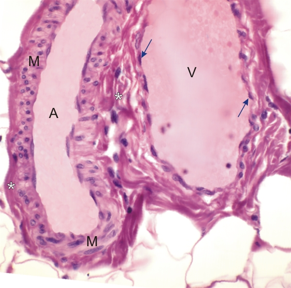
Longitudinal sections of an arteriole (A) and a venule (V).

The arteriole on the left does not show an internal elastic lamella under the endothelium.

The media (M) is composed of two layers of smooth muscle cells seen here in transverse or oblique sections. The mediae of larger vessels are composed of three or more layers of smooth muscle cells, as in the arteries shown in figures 5.15 to 5.21.

The adventitia (*) is thin and merges, on the right side, with the connective tissue surrounding the venule.

The thin wall of the venule (on the right) is composed of an endothelium (arrows) lining some dense connective tissue, free of smooth muscle fibres.

Stain: H–­­E
Magnification: ×600


We'd very much appreciate receiving your comments and suggestions!We'd very much appreciate receiving your comments and suggestions!

Creative Commons Licence The text and images of this Histology Atlas, by Yves Clermont, Michael Lalli & Zsuzsanna Bencsath-Makkai, are licensed under a Creative Commons Attribution-Noncommercial-No Derivative Works 2.5 Canada Licence and cannot be modified without the written permission of the authors. Use of any text or images must carry an acknowledgement which includes a link to the original work.


HAweb/Zs B-M -> HAbase-generated on 2013-09-13