Contents Yves Clermont, Michael Lalli, Zsuzsanna Bencsath-Makkai
LIGHT-MICROSCOPIC HISTOLOGY ATLAS
     5. Vessels
  1     2     3     4     5     6     7     8     9     10     11     12     13     14     15     16     17     18     19     20     21     22     23     24     25  
  26     27     28     29     30     31     32     33     34     35  

    5.8   Artery Version française
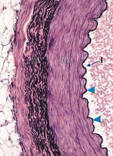
Transverse section of a large artery stained with Verhoeff’s method.

This vessel shows three distinct layers, or tunica: the intima (I), the media (M) and the adventitia (A).

The intima is composed of an endothelium (difficult to see at this magnification) applied to the internal elastic lamella (arrowhead).

The media is composed of circularly arranged smooth muscle cells and of a few elastic fibres.

The thick adventitia does not contain muscle cells but is composed of elastic fibres and lamellae (black) and collagen fibres (pink).

Stain: Verhoeff
Magnification: ×250


We'd very much appreciate receiving your comments and suggestions!We'd very much appreciate receiving your comments and suggestions!

Creative Commons Licence The text and images of this Histology Atlas, by Yves Clermont, Michael Lalli & Zsuzsanna Bencsath-Makkai, are licensed under a Creative Commons Attribution-Noncommercial-No Derivative Works 2.5 Canada Licence and cannot be modified without the written permission of the authors. Use of any text or images must carry an acknowledgement which includes a link to the original work.


HAweb/Zs B-M -> HAbase-generated on 2013-09-13