Contents Yves Clermont, Michael Lalli, Zsuzsanna Bencsath-Makkai
LIGHT-MICROSCOPIC HISTOLOGY ATLAS
     5. Vessels
  1     2     3     4     5     6     7     8     9     10     11     12     13     14     15     16     17     18     19     20     21     22     23     24     25  
  26     27     28     29     30     31     32     33     34     35  

    5.27   Vessels Version française
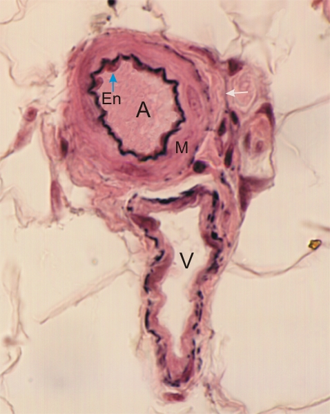
Transverse thick sections of a small artery (A) and of an accompanying small vein (V). This is a higher magnification of the framed area in Figure 5.14.

The artery shows an intima with its endothelium (En) and a well-stained internal elastic lamina.

The media (M) is composed mainly of smooth muscle cells.

Its thin adventitia (arrow) includes collagen and a few elastic fibres.

The accompanying venule does not show a clear-cut endothelium and its wall is composed of collagen fibres (pink) and a few elastic fibres (black).

Stain: Verhoeff
Magnification: ×1000


We'd very much appreciate receiving your comments and suggestions!We'd very much appreciate receiving your comments and suggestions!

Creative Commons Licence The text and images of this Histology Atlas, by Yves Clermont, Michael Lalli & Zsuzsanna Bencsath-Makkai, are licensed under a Creative Commons Attribution-Noncommercial-No Derivative Works 2.5 Canada Licence and cannot be modified without the written permission of the authors. Use of any text or images must carry an acknowledgement which includes a link to the original work.


HAweb/Zs B-M -> HAbase-generated on 2013-09-13