Contents Yves Clermont, Michael Lalli, Zsuzsanna Bencsath-Makkai
LIGHT-MICROSCOPIC HISTOLOGY ATLAS
     5. Vessels
  1     2     3     4     5     6     7     8     9     10     11     12     13     14     15     16     17     18     19     20     21     22     23     24     25  
  26     27     28     29     30     31     32     33     34     35  

    5.20   Artery Version française
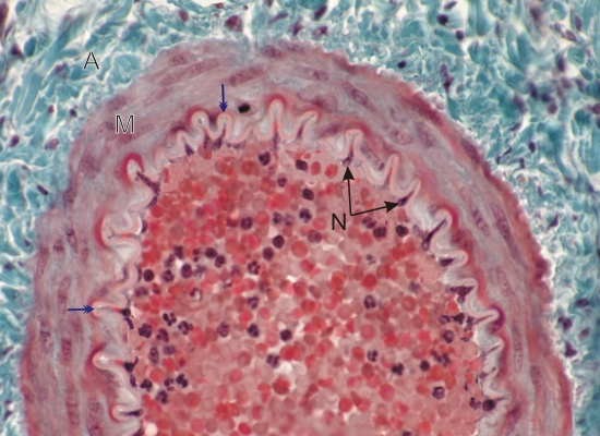
Transverse section of a small artery.

The lumen of the vessel contains red blood cells and several basophilic leukocytes.

Inside the unstained wavy internal elastic lamella (blue arrows), the nuclei of endothelial cells (N) are seen in depressions of the intima. These folds are artefacts due to the contraction of the tissues during fixation.

The media (M) shows three to five concentric layers of smooth muscle cells.

The adventitia (A), composed of dense connective tissue, is bright blue-green in this preparation.

Stain: Masson’s Trichrome
Magnification: ×650


We'd very much appreciate receiving your comments and suggestions!We'd very much appreciate receiving your comments and suggestions!

Creative Commons Licence The text and images of this Histology Atlas, by Yves Clermont, Michael Lalli & Zsuzsanna Bencsath-Makkai, are licensed under a Creative Commons Attribution-Noncommercial-No Derivative Works 2.5 Canada Licence and cannot be modified without the written permission of the authors. Use of any text or images must carry an acknowledgement which includes a link to the original work.


HAweb/Zs B-M -> HAbase-generated on 2013-09-13