Contents Yves Clermont, Michael Lalli, Zsuzsanna Bencsath-Makkai
LIGHT-MICROSCOPIC HISTOLOGY ATLAS
     5. Vessels
  1     2     3     4     5     6     7     8     9     10     11     12     13     14     15     16     17     18     19     20     21     22     23     24     25  
  26     27     28     29     30     31     32     33     34     35  

    5.25   Arterioles and venule Version française
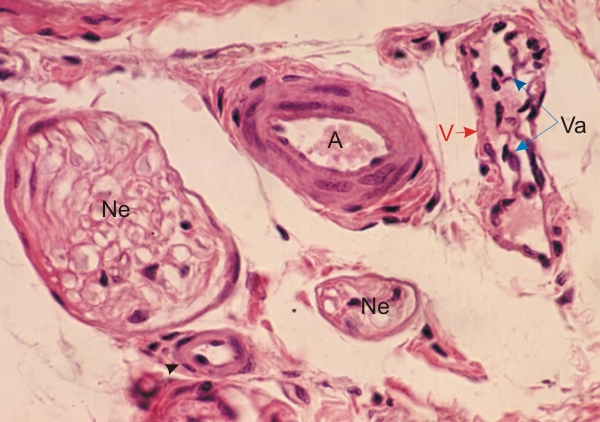
Section of a cat tongue.

This field shows cross sections of a large arteriole (A), a small arteriole (arrowhead), large and small nerves (Ne) and a venule (V) with valves (Va).

The large arteriole shows two concentric layers of smooth muscle cells. The small arteriole shows a single layer of smooth muscle cells. In both vessels the adventitia is limited to a small amount of connective tissue.

Stain: H–­­E
Magnification: ×450


We'd very much appreciate receiving your comments and suggestions!We'd very much appreciate receiving your comments and suggestions!

Creative Commons Licence The text and images of this Histology Atlas, by Yves Clermont, Michael Lalli & Zsuzsanna Bencsath-Makkai, are licensed under a Creative Commons Attribution-Noncommercial-No Derivative Works 2.5 Canada Licence and cannot be modified without the written permission of the authors. Use of any text or images must carry an acknowledgement which includes a link to the original work.


HAweb/Zs B-M -> HAbase-generated on 2013-09-13