Contents Yves Clermont, Michael Lalli, Zsuzsanna Bencsath-Makkai
LIGHT-MICROSCOPIC HISTOLOGY ATLAS
     5. Vessels
  1     2     3     4     5     6     7     8     9     10     11     12     13     14     15     16     17     18     19     20     21     22     23     24     25  
  26     27     28     29     30     31     32     33     34     35  

    5.14   Vein Version française
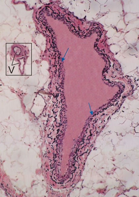
Transverse section of a medium-sized vein.

Note that the irregular lumen of the vein is wide and its wall relatively thin. There is no internal elastic lamella in the wall of this vein. The purplish layer underlying the endothelium contains some smooth muscle cells plus collagen and elastic fibres (blue arrows). The external layer of the vein contains acidophilic collagen and black elastic fibres.

(Framed area) In the loose connective tissue surrounding the vein there are two small vessels (V) shown at a higher magnification in Figure 5.27.

Stain: Verhoeff
Magnification: ×150


We'd very much appreciate receiving your comments and suggestions!We'd very much appreciate receiving your comments and suggestions!

Creative Commons Licence The text and images of this Histology Atlas, by Yves Clermont, Michael Lalli & Zsuzsanna Bencsath-Makkai, are licensed under a Creative Commons Attribution-Noncommercial-No Derivative Works 2.5 Canada Licence and cannot be modified without the written permission of the authors. Use of any text or images must carry an acknowledgement which includes a link to the original work.


HAweb/Zs B-M -> HAbase-generated on 2013-09-13