|
||
| 5. Vessels | ||
| 1 2 3 4 5 6 7 8 9 10 11 12 13 14 15 16 17 18 19 20 21 22 23 24 25 | ||
| 26 27 28 29 30 31 32 33 34 35 |
| |||
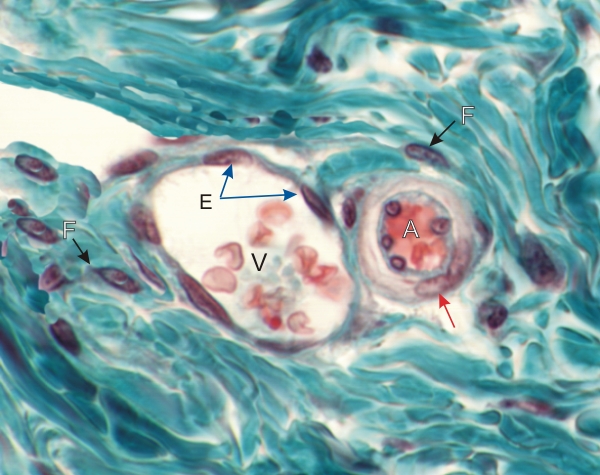 |
In this field the dense connective tissue is stained green. It includes two transverse sections of small vessels, a small arteriole (A) and a venule (V).
The arteriole shows the nuclei of endothelial cells of the intima and a single layer of smooth muscle cells (arrow) in the media. The accompanying venule has a large lumen containing red blood cells and a thin wall showing endothelial cells (E) surrounded by connective tissue. The nuclei of fibrocytes (F) present in the surrounding connective tissue are also labelled. Stain: Massons Trichrome
|
||