|
||
| 5. Vessels | ||
| 1 2 3 4 5 6 7 8 9 10 11 12 13 14 15 16 17 18 19 20 21 22 23 24 25 | ||
| 26 27 28 29 30 31 32 33 34 35 |
| |||
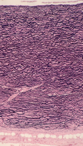 |
Transverse section of the aorta to show the elastic lamellae.
Lining the lumen (top), a thin indistinct layer corresponds to the intima. Most of the wall is composed of superposed elastic lamellae forming the media. These elastic lamellae are separated by an acidophilic material composed of smooth muscle cells and collagen. The acidophilic layer (bottom) is composed mainly of collagen that forms a thin adventitia. Stain: Verhoeff
|
||