Contents Yves Clermont, Michael Lalli, Zsuzsanna Bencsath-Makkai
LIGHT-MICROSCOPIC HISTOLOGY ATLAS
     5. Vessels
  1     2     3     4     5     6     7     8     9     10     11     12     13     14     15     16     17     18     19     20     21     22     23     24     25  
  26     27     28     29     30     31     32     33     34     35  

    5.10   Artery Version française
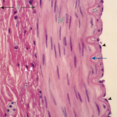
Transverse section of a medium-sized artery.

The intima shows the endothelium (arrowheads) separated from the acidophilic internal elastic lamella (blue arrow) by a layer of connective tissue (*).

The media (M) is composed of smooth muscle cells, which are circularly arranged and show their elongated nuclei.

The adventitia (A) shows a dense connective tissue, in which collagen and elastin, both acidophilic, cannot be differentiated. The nuclei of fibrocytes (F) are indicated in this adventitia.

Stain: H–­­E
Magnification: ×500


We'd very much appreciate receiving your comments and suggestions!We'd very much appreciate receiving your comments and suggestions!

Creative Commons Licence The text and images of this Histology Atlas, by Yves Clermont, Michael Lalli & Zsuzsanna Bencsath-Makkai, are licensed under a Creative Commons Attribution-Noncommercial-No Derivative Works 2.5 Canada Licence and cannot be modified without the written permission of the authors. Use of any text or images must carry an acknowledgement which includes a link to the original work.


HAweb/Zs B-M -> HAbase-generated on 2013-09-13