|
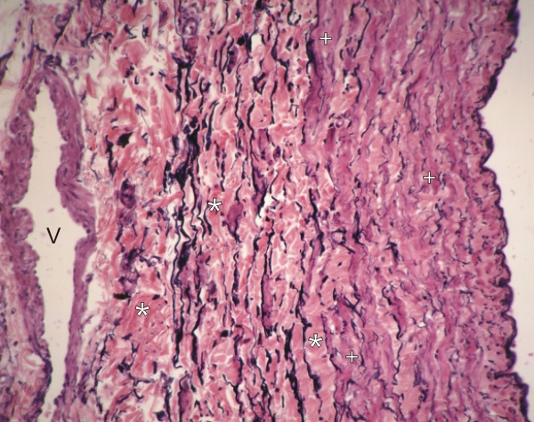
Section through the wall of a large vein.
The intima is on the right and the adventitia is on the left. The greater part of the wall of this vessel is composed of irregular bundles of purple-pink smooth muscle cells (+) separated by an appreciable amount of pink collagen (*) and elastic fibres (black). The walls of the veins are less well organized than those of the arteries, and the demarcations of the intima, media and adventitia are not clear.
A small vein (V) is visible in the external wall of this large vein.
Stain: Verhoeff
Magnification: ×200
|