Contents Yves Clermont, Michael Lalli, Zsuzsanna Bencsath-Makkai
LIGHT-MICROSCOPIC HISTOLOGY ATLAS
     5. Vessels
  1     2     3     4     5     6     7     8     9     10     11     12     13     14     15     16     17     18     19     20     21     22     23     24     25  
  26     27     28     29     30     31     32     33     34     35  

    5.15   Artery Version française
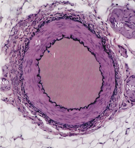
Transverse section of a medium-sized artery.

The three tunicae forming the wall of this vessel are distinct. The intima (I) shows the wavy internal elastic lamella. The media (M) is composed of smooth muscle cells and of very few elastic fibres. The adventitia is a layer of dense connective tissue composed of collagen and elastic fibres.

This vessel is surrounded by loose connective tissue containing adipocytes and three cross sections of nerves (Ne).

Stain: Verhoeff
Magnification: ×200


We'd very much appreciate receiving your comments and suggestions!We'd very much appreciate receiving your comments and suggestions!

Creative Commons Licence The text and images of this Histology Atlas, by Yves Clermont, Michael Lalli & Zsuzsanna Bencsath-Makkai, are licensed under a Creative Commons Attribution-Noncommercial-No Derivative Works 2.5 Canada Licence and cannot be modified without the written permission of the authors. Use of any text or images must carry an acknowledgement which includes a link to the original work.


HAweb/Zs B-M -> HAbase-generated on 2013-09-13