Contents Yves Clermont, Michael Lalli, Zsuzsanna Bencsath-Makkai
LIGHT-MICROSCOPIC HISTOLOGY ATLAS
     5. Vessels
  1     2     3     4     5     6     7     8     9     10     11     12     13     14     15     16     17     18     19     20     21     22     23     24     25  
  26     27     28     29     30     31     32     33     34     35  

    5.3   Aorta Version française
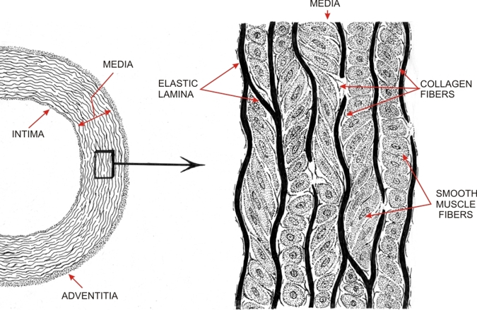
The drawing is a schematic representation of the aorta, an elastic artery.

The framed area of the media is shown on the right to illustrate the disposition of the smooth muscle cells between the fenestrated elastic lamellae. These muscle cells are short and their pointed extremities attach to the surfaces of two adjacent elastic lamellae. These smooth muscle cells are arranged in groups which have different orientations as indicated by their longitudinal, oblique and cross-sectional profiles.

The presence of numerous elastic lamellae and of smooth muscle cells attached to these lamellae contribute to the solidity and resilience of the wall of these arteries submitted to the strong rhythmic pulsations of the heart.


We'd very much appreciate receiving your comments and suggestions!We'd very much appreciate receiving your comments and suggestions!

Creative Commons Licence The text and images of this Histology Atlas, by Yves Clermont, Michael Lalli & Zsuzsanna Bencsath-Makkai, are licensed under a Creative Commons Attribution-Noncommercial-No Derivative Works 2.5 Canada Licence and cannot be modified without the written permission of the authors. Use of any text or images must carry an acknowledgement which includes a link to the original work.


HAweb/Zs B-M -> HAbase-generated on 2013-09-13