Sommaire Yves Clermont, Michael Lalli, Zsuzsanna Bencsath-Makkai
ATLAS D'HISTOLOGIE EN MICROSCOPIE OPTIQUE
     5. Vaisseaux
  1     2     3     4     5     6     7     8     9     10     11     12     13     14     15     16     17     18     19     20     21     22     23     24     25  
  26     27     28     29     30     31     32     33     34     35  
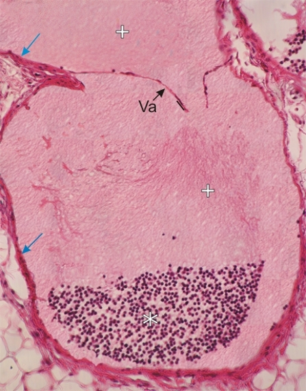
    5.34   Vaisseau lymphatique English version
Coupe d’un gros vaisseau lymphatique de la cavité thoracique.

Ce champ montre une valve (Va) formée de deux minces feuillets formés par un endothélium posé sur un fine lame conjonctive. La lymphe (+) est présente des deux cotés de la valve. Au bas du champ on remarque l’accumulation d’un grand nombre de lymphocytes (*).

La paroi conjonctive du vaisseau est également indiquée (flèches).

Coloration: H–­­É
Grossissement: ×200


We'd very much appreciate receiving your comments and suggestions!We'd very much appreciate receiving your comments and suggestions!

Creative Commons Licence The text and images of this Histology Atlas, by Yves Clermont, Michael Lalli & Zsuzsanna Bencsath-Makkai, are licensed under a Creative Commons Attribution-Noncommercial-No Derivative Works 2.5 Canada Licence and cannot be modified without the written permission of the authors. Use of any text or images must carry an acknowledgement which includes a link to the original work.


HAweb/Zs B-M -> HAbase-generated on 2013-09-13