Contents Yves Clermont, Michael Lalli, Zsuzsanna Bencsath-Makkai
LIGHT-MICROSCOPIC HISTOLOGY ATLAS
     5. Vessels
  1     2     3     4     5     6     7     8     9     10     11     12     13     14     15     16     17     18     19     20     21     22     23     24     25  
  26     27     28     29     30     31     32     33     34     35  

    5.35   Lymphatic vessel Version française
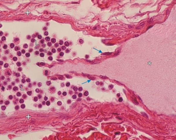
Section of a small lymphatic vessel.

This field shows a longitudinal section of the vessel at the level of a valvule with its two leaflets. These two thin membranes are lined with an endothelium (arrows). In the lumen of the vessel on the right, the lymph (*) is free of cells. On the left, the lumen of the vessel contains numerous lymphocytes (L).

Obviously, the valve prevents the retrograde flow of these lymphocytes along the vessel. The connective tissue of the lateral wall is indicated (+).

Stain: H–­­E
Magnification: ×750


We'd very much appreciate receiving your comments and suggestions!We'd very much appreciate receiving your comments and suggestions!

Creative Commons Licence The text and images of this Histology Atlas, by Yves Clermont, Michael Lalli & Zsuzsanna Bencsath-Makkai, are licensed under a Creative Commons Attribution-Noncommercial-No Derivative Works 2.5 Canada Licence and cannot be modified without the written permission of the authors. Use of any text or images must carry an acknowledgement which includes a link to the original work.


HAweb/Zs B-M -> HAbase-generated on 2013-09-13