Contents Yves Clermont, Michael Lalli, Zsuzsanna Bencsath-Makkai
LIGHT-MICROSCOPIC HISTOLOGY ATLAS
     9. Skin
  1     2     3     4     5     6     7     8     9     10     11     12     13     14     15     16     17     18     19     20     21     22     23     24     25  
  26     27     28     29     30     31     32     33     34     35  

    9.7   Thin skin Version française
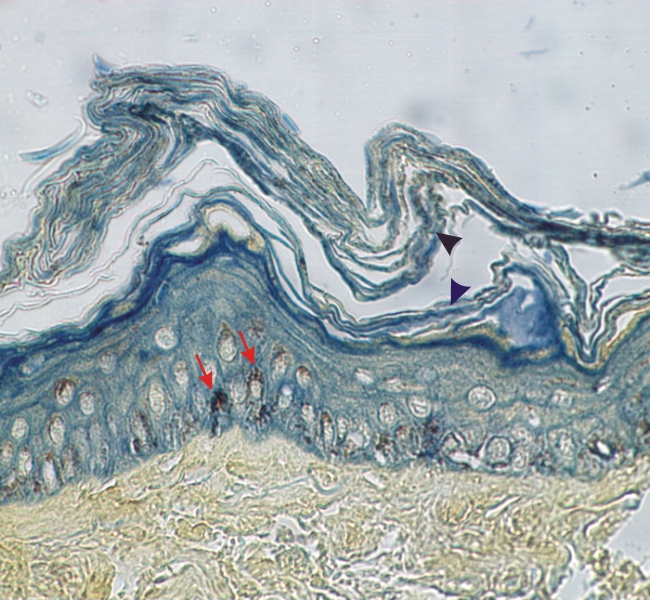
Section of thin skin stained with TPA.

In this section the nuclei of keratinocytes are pale but the cytoplasms are lightly stained green owing to the presence of keratin filaments. The brown staining of the cytoplasm of keratinocytes is due to melanin granules (arrows).

The desquamating flat cell residues (arrowheads) of the cornified layer are well stained with TPA.

The subjacent connective tissue is stained light yellow.

Stain: TPA
Magnification: ×900


We'd very much appreciate receiving your comments and suggestions!We'd very much appreciate receiving your comments and suggestions!

Creative Commons Licence The text and images of this Histology Atlas, by Yves Clermont, Michael Lalli & Zsuzsanna Bencsath-Makkai, are licensed under a Creative Commons Attribution-Noncommercial-No Derivative Works 2.5 Canada Licence and cannot be modified without the written permission of the authors. Use of any text or images must carry an acknowledgement which includes a link to the original work.


HAweb/Zs B-M -> HAbase-generated on 2013-09-13