Contents Yves Clermont, Michael Lalli, Zsuzsanna Bencsath-Makkai
LIGHT-MICROSCOPIC HISTOLOGY ATLAS
     9. Skin
  1     2     3     4     5     6     7     8     9     10     11     12     13     14     15     16     17     18     19     20     21     22     23     24     25  
  26     27     28     29     30     31     32     33     34     35  

    9.25   Hair formation Version française
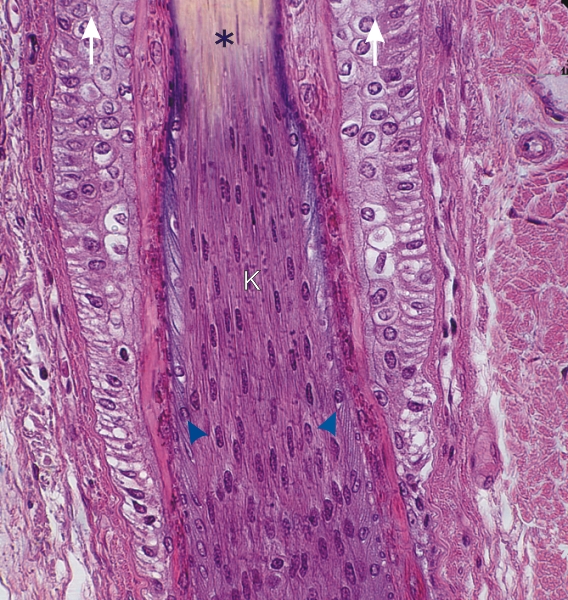
This field corresponds to the framed area of Figure 9.24. It shows the keratogenous zone of a growing hair.

The external root sheath (vertical arrows) of the follicle decreases in thickness as it approaches the bulb. The fully formed hair (*), composed of the hard keratin of the cortex, derives from the elongated cells of the keratogenous zone (K). These elongated cells derive from the matrix cells of the bulb and undergo keratinization without the formation of keratohyalin granules.

At the periphery of the keratogenous zone, the flattened basophilic cells (arrowheads) are the precursors of the cuticle of the hair. These are separated from the external root sheath by the acidophilic material of the internal root sheath.

Stain: H–­­E
Magnification: ×750


We'd very much appreciate receiving your comments and suggestions!We'd very much appreciate receiving your comments and suggestions!

Creative Commons Licence The text and images of this Histology Atlas, by Yves Clermont, Michael Lalli & Zsuzsanna Bencsath-Makkai, are licensed under a Creative Commons Attribution-Noncommercial-No Derivative Works 2.5 Canada Licence and cannot be modified without the written permission of the authors. Use of any text or images must carry an acknowledgement which includes a link to the original work.


HAweb/Zs B-M -> HAbase-generated on 2013-09-13