Contents Yves Clermont, Michael Lalli, Zsuzsanna Bencsath-Makkai
LIGHT-MICROSCOPIC HISTOLOGY ATLAS
     9. Skin
  1     2     3     4     5     6     7     8     9     10     11     12     13     14     15     16     17     18     19     20     21     22     23     24     25  
  26     27     28     29     30     31     32     33     34     35  

    9.29   Hair follicle Version française
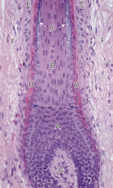
Section of the root of a hair follicle.

This field shows the upper part of the bulb and the proximal segment of the keratogenous zone.

The following structures are labelled:

  • Matrix cells of the bulb (A)
  • Elongated cells of the keratogenous zone (B). Note the sharp demarcation (horizontal arrows and the stippled line) between A and B.
  • Single layer of flattened basophilic cells destined to form the cuticle of the hair (C)
  • Acidophilic internal root sheath (D)
  • External root sheath (E)
  • Connective tissue sheath around the hair follicle (F)
  • Papilla of the bulb (G)

Stain: H–­­E
Magnification: ×750


We'd very much appreciate receiving your comments and suggestions!We'd very much appreciate receiving your comments and suggestions!

Creative Commons Licence The text and images of this Histology Atlas, by Yves Clermont, Michael Lalli & Zsuzsanna Bencsath-Makkai, are licensed under a Creative Commons Attribution-Noncommercial-No Derivative Works 2.5 Canada Licence and cannot be modified without the written permission of the authors. Use of any text or images must carry an acknowledgement which includes a link to the original work.


HAweb/Zs B-M -> HAbase-generated on 2013-09-13