Contents Yves Clermont, Michael Lalli, Zsuzsanna Bencsath-Makkai
LIGHT-MICROSCOPIC HISTOLOGY ATLAS
     9. Skin
  1     2     3     4     5     6     7     8     9     10     11     12     13     14     15     16     17     18     19     20     21     22     23     24     25  
  26     27     28     29     30     31     32     33     34     35  

    9.1   Thick skin Version française
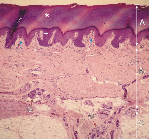
Section of the thick skin of the palm of a hand.

The three main layers of the skin are visible:

  • Epidermis (A) with its thick superficial cornified layer (stratum corneum) (*) and its underlying stratified squamous epithelium (+).
  • Dermis (B) mainly composed of dense connective tissue (including vessels and nerves) showing dermal papillae (vertical arrows) which project into the epithelium.
  • Hypodermis (C) composed of areas of adipose tissue separated by septae of dense connective tissue.

Profiles of sweat glands (G) are seen at the borderline of the dermis and hypodermis. On the right, the duct of a sweat gland crosses the dermis in the direction of the surface of the skin.

Stain: H–­­E
Magnification: ×100


We'd very much appreciate receiving your comments and suggestions!We'd very much appreciate receiving your comments and suggestions!

Creative Commons Licence The text and images of this Histology Atlas, by Yves Clermont, Michael Lalli & Zsuzsanna Bencsath-Makkai, are licensed under a Creative Commons Attribution-Noncommercial-No Derivative Works 2.5 Canada Licence and cannot be modified without the written permission of the authors. Use of any text or images must carry an acknowledgement which includes a link to the original work.


HAweb/Zs B-M -> HAbase-generated on 2013-09-13