Contents Yves Clermont, Michael Lalli, Zsuzsanna Bencsath-Makkai
LIGHT-MICROSCOPIC HISTOLOGY ATLAS
     9. Skin
  1     2     3     4     5     6     7     8     9     10     11     12     13     14     15     16     17     18     19     20     21     22     23     24     25  
  26     27     28     29     30     31     32     33     34     35  

    9.30   Hair follicle Version française
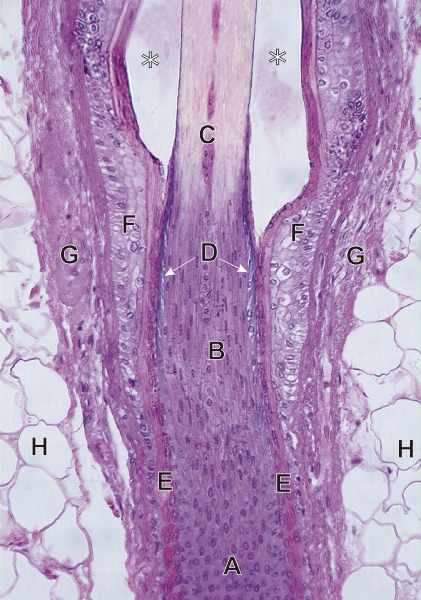
Section of a hair follicle.

This field shows the following features:

  • Epithelial cells of the hair matrix (A)
  • Transformation of epithelial cells into the elongated cells of the keratogenous zone (B)
  • Transformation of the elongated cells (C) into hard keratin (pale yellow) of the hair cortex. This hair shows a narrow medulla in the centre of the cortex.
  • Layer of basophilic cells that form the cuticle (D)
  • Acidophilic inner root sheath which disintegrates along the keratogenous zone (E)
  • External root sheath (F)
  • Connective tissue sheath of the follicle (G)
  • Adipose tissue of the hypodermis (H)

The spaces (*) seen above between the hair and the external root sheath are artefacts of tissue preparation.

Stain: H–­­E
Magnification: ×325


We'd very much appreciate receiving your comments and suggestions!We'd very much appreciate receiving your comments and suggestions!

Creative Commons Licence The text and images of this Histology Atlas, by Yves Clermont, Michael Lalli & Zsuzsanna Bencsath-Makkai, are licensed under a Creative Commons Attribution-Noncommercial-No Derivative Works 2.5 Canada Licence and cannot be modified without the written permission of the authors. Use of any text or images must carry an acknowledgement which includes a link to the original work.


HAweb/Zs B-M -> HAbase-generated on 2013-09-13