Contents Yves Clermont, Michael Lalli, Zsuzsanna Bencsath-Makkai
LIGHT-MICROSCOPIC HISTOLOGY ATLAS
     9. Skin
  1     2     3     4     5     6     7     8     9     10     11     12     13     14     15     16     17     18     19     20     21     22     23     24     25  
  26     27     28     29     30     31     32     33     34     35  

    9.3   Epidermis of thick skin. Version française

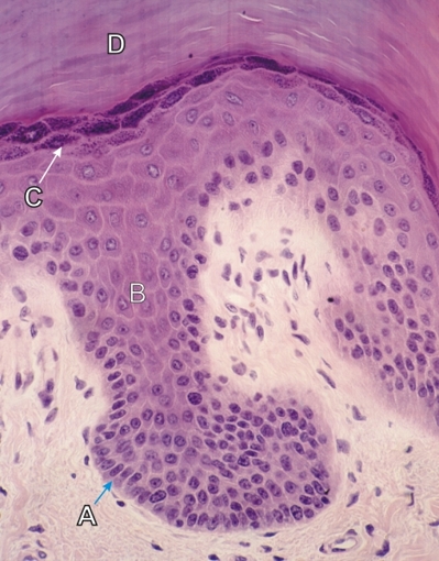
The following layers of the epithelium are labelled:

  • Basal cell layer (stratum basale) (A) in which the epithelial cells divide. They renew themselves and give rise to the epithelial cells of the stratum spinosum above.
  • Polyhedral cell layer (stratum spinosum) (B). These cells, which show pale boundaries, transform into the cells of the next layer above.
  • Granular cell layer (stratum granulosum) (C) composed of squamous cells containing an increasing number of basophilic keratohyalin granules.
  • Cornified layer (stratum corneum) (D) formed of epithelial cell residues derived from the sudden transformation of the cells of the stratum granulosum. This layer is composed of soft keratin.

All the nucleated epithelial cells of this epithelium may be called keratinocytes.

Stain: H–­­E
Magnification: ×900


We'd very much appreciate receiving your comments and suggestions!We'd very much appreciate receiving your comments and suggestions!

Creative Commons Licence The text and images of this Histology Atlas, by Yves Clermont, Michael Lalli & Zsuzsanna Bencsath-Makkai, are licensed under a Creative Commons Attribution-Noncommercial-No Derivative Works 2.5 Canada Licence and cannot be modified without the written permission of the authors. Use of any text or images must carry an acknowledgement which includes a link to the original work.


HAweb/Zs B-M -> HAbase-generated on 2013-09-13