Contents Yves Clermont, Michael Lalli, Zsuzsanna Bencsath-Makkai
LIGHT-MICROSCOPIC HISTOLOGY ATLAS
     9. Skin
  1     2     3     4     5     6     7     8     9     10     11     12     13     14     15     16     17     18     19     20     21     22     23     24     25  
  26     27     28     29     30     31     32     33     34     35  

    9.19   Hair follicle and sebaceous glands Version française
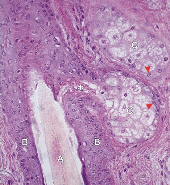
Section of the neck of a hair follicle.

The following components are labelled:

  • Pale stained hair shaft (A)
  • External root sheath (B)
  • Two sebaceous glands (C), one with an excretory duct (*) opening into the space around the hair.

The sebaceous glands show their basal cells (arrowheads) and their lipid-containing daughter cells. In the sebaceous gland some epithelial cells degenerate (arrows) as they reach the excretory duct.

Stain: H–­­E
Magnification: ×900


We'd very much appreciate receiving your comments and suggestions!We'd very much appreciate receiving your comments and suggestions!

Creative Commons Licence The text and images of this Histology Atlas, by Yves Clermont, Michael Lalli & Zsuzsanna Bencsath-Makkai, are licensed under a Creative Commons Attribution-Noncommercial-No Derivative Works 2.5 Canada Licence and cannot be modified without the written permission of the authors. Use of any text or images must carry an acknowledgement which includes a link to the original work.


HAweb/Zs B-M -> HAbase-generated on 2013-09-13