Contents Yves Clermont, Michael Lalli, Zsuzsanna Bencsath-Makkai
LIGHT-MICROSCOPIC HISTOLOGY ATLAS
     9. Skin
  1     2     3     4     5     6     7     8     9     10     11     12     13     14     15     16     17     18     19     20     21     22     23     24     25  
  26     27     28     29     30     31     32     33     34     35  

    9.26   Hair bulb Version française
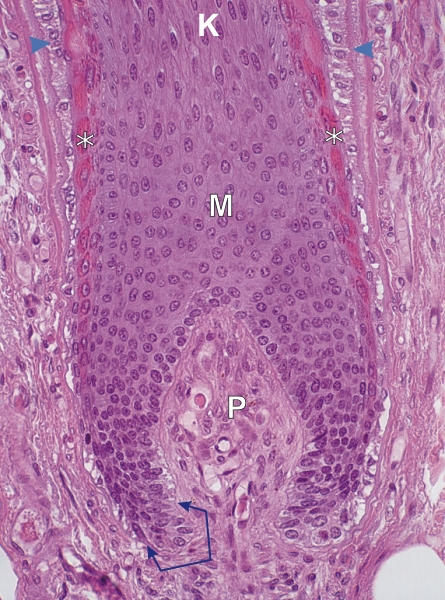
Longitudinal section of a hair bulb.

The connective tissue papilla (P) occupies the centre of the bulb and is surrounded by the matrix of the bulb (M).

This matrix is composed of epithelial cells derived from a layer of undifferentiated basal cells (arrows). This layer of dividing and renewing basal cells is continuous with the basal cell layer of the external root sheath (arrowheads above).

The acidophilic cell layers (*) belong to the internal root sheath surrounding the bulb which has a transitory existence (see Figure 9.23).

Most of the matrix cells elongate above as they enter the keratogenous zone (K).

Stain: H–­­E
Magnification: ×750


We'd very much appreciate receiving your comments and suggestions!We'd very much appreciate receiving your comments and suggestions!

Creative Commons Licence The text and images of this Histology Atlas, by Yves Clermont, Michael Lalli & Zsuzsanna Bencsath-Makkai, are licensed under a Creative Commons Attribution-Noncommercial-No Derivative Works 2.5 Canada Licence and cannot be modified without the written permission of the authors. Use of any text or images must carry an acknowledgement which includes a link to the original work.


HAweb/Zs B-M -> HAbase-generated on 2013-09-13