Contents Yves Clermont, Michael Lalli, Zsuzsanna Bencsath-Makkai
LIGHT-MICROSCOPIC HISTOLOGY ATLAS
     9. Skin
  1     2     3     4     5     6     7     8     9     10     11     12     13     14     15     16     17     18     19     20     21     22     23     24     25  
  26     27     28     29     30     31     32     33     34     35  

    9.20   Hair follicle and sebaceous glands Version française
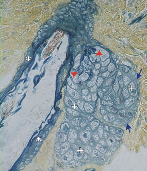
Hair follicle and an associated sebaceous gland stained with TPA.

This field shows the detail of a sebaceous gland with a few basal cells (arrows) and lipid -containing cells (+). The latter are sharply demarcated and show a central nucleus surrounded by a vacuolated cytoplasm. These vacuoles are spaces occupied by lipid droplets which were dissolved during the preparation of the tissue section. These cells degenerate as they are extruded into the excretory duct (arrowheads).

The external root sheath of the follicle is also indicated (*).

Stain: TPA
Magnification: ×900


We'd very much appreciate receiving your comments and suggestions!We'd very much appreciate receiving your comments and suggestions!

Creative Commons Licence The text and images of this Histology Atlas, by Yves Clermont, Michael Lalli & Zsuzsanna Bencsath-Makkai, are licensed under a Creative Commons Attribution-Noncommercial-No Derivative Works 2.5 Canada Licence and cannot be modified without the written permission of the authors. Use of any text or images must carry an acknowledgement which includes a link to the original work.


HAweb/Zs B-M -> HAbase-generated on 2013-09-13