Contents Yves Clermont, Michael Lalli, Zsuzsanna Bencsath-Makkai
LIGHT-MICROSCOPIC HISTOLOGY ATLAS
     9. Skin
  1     2     3     4     5     6     7     8     9     10     11     12     13     14     15     16     17     18     19     20     21     22     23     24     25  
  26     27     28     29     30     31     32     33     34     35  

    9.10   Sensory corpuscle of Pacini Version française
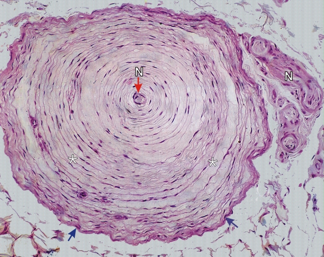
Section of the hypodermis of a toe showing a sensory corpuscle of Pacini.

This corpuscle consists of numerous (30–35) concentric lamellae (*) composed of flattened fibrocytes separated by spaces containing few fine connective tissue fibres and a fluid of low viscosity.

This corpuscle is delimited by a connective tissue capsule (arrows). The nerve (N) enters the corpuscle at one pole and sends an axon toward its centre.

This sensory organ permits the discrimination of variations in pressure applied to the skin.

Stain: H–­­E
Magnification: ×300


We'd very much appreciate receiving your comments and suggestions!We'd very much appreciate receiving your comments and suggestions!

Creative Commons Licence The text and images of this Histology Atlas, by Yves Clermont, Michael Lalli & Zsuzsanna Bencsath-Makkai, are licensed under a Creative Commons Attribution-Noncommercial-No Derivative Works 2.5 Canada Licence and cannot be modified without the written permission of the authors. Use of any text or images must carry an acknowledgement which includes a link to the original work.


HAweb/Zs B-M -> HAbase-generated on 2013-09-13