Contents Yves Clermont, Michael Lalli, Zsuzsanna Bencsath-Makkai
LIGHT-MICROSCOPIC HISTOLOGY ATLAS
     9. Skin
  1     2     3     4     5     6     7     8     9     10     11     12     13     14     15     16     17     18     19     20     21     22     23     24     25  
  26     27     28     29     30     31     32     33     34     35  

    9.4   Thick skin Version française
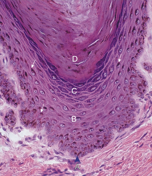
Section of thick skin.

This field is similar to that of Figure 9.3, but the keratinocytes of the basal and polyhedral cell layers contain pigment granules of melanin (arrows). These brown granules accumulate in the cytoplasm next to the nuclei. Located among the basal cells, the melanocytes, which produce the pigment granules, cannot be identified in this section. These melanin granules, once secreted by the melanocytes, are endocytosed by the keratinocytes.

The following cell layers of the epidermis are labelled:

  • Basal layer, or stratum basale (A)
  • Polyhedral cell layer, or stratum spinosum (B)
  • Granular cell layer, or stratum granulosum (C)
  • Cornified cell layer, or stratum corneum (D)

Stain: H–­­E
Magnification: ×900


We'd very much appreciate receiving your comments and suggestions!We'd very much appreciate receiving your comments and suggestions!

Creative Commons Licence The text and images of this Histology Atlas, by Yves Clermont, Michael Lalli & Zsuzsanna Bencsath-Makkai, are licensed under a Creative Commons Attribution-Noncommercial-No Derivative Works 2.5 Canada Licence and cannot be modified without the written permission of the authors. Use of any text or images must carry an acknowledgement which includes a link to the original work.


HAweb/Zs B-M -> HAbase-generated on 2013-09-13