Contents Yves Clermont, Michael Lalli, Zsuzsanna Bencsath-Makkai
LIGHT-MICROSCOPIC HISTOLOGY ATLAS
     9. Skin
  1     2     3     4     5     6     7     8     9     10     11     12     13     14     15     16     17     18     19     20     21     22     23     24     25  
  26     27     28     29     30     31     32     33     34     35  

    9.2   Thick skin Version française
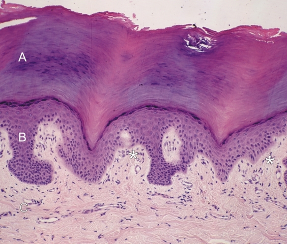
Section of the epidermis of thick skin.

This field shows the two main layers of the epidermis: the stratum corneum (A) and the stratified squamous epithelium (B).

The cornified layer of the epidermis is composed of soft keratin formed by the accumulation of cytoskeletal residues derived from the underlying squamous epidermal cells.

The dermis (C) is composed of a dense connective tissue. The dermal papillae (*) of this tissue deeply invaginate the internal surface of the epidermis.

Stain: H–­­E
Magnification: ×225


We'd very much appreciate receiving your comments and suggestions!We'd very much appreciate receiving your comments and suggestions!

Creative Commons Licence The text and images of this Histology Atlas, by Yves Clermont, Michael Lalli & Zsuzsanna Bencsath-Makkai, are licensed under a Creative Commons Attribution-Noncommercial-No Derivative Works 2.5 Canada Licence and cannot be modified without the written permission of the authors. Use of any text or images must carry an acknowledgement which includes a link to the original work.


HAweb/Zs B-M -> HAbase-generated on 2013-09-13