Contents Yves Clermont, Michael Lalli, Zsuzsanna Bencsath-Makkai
LIGHT-MICROSCOPIC HISTOLOGY ATLAS
     9. Skin
  1     2     3     4     5     6     7     8     9     10     11     12     13     14     15     16     17     18     19     20     21     22     23     24     25  
  26     27     28     29     30     31     32     33     34     35  

    9.5   Epidermis Version française
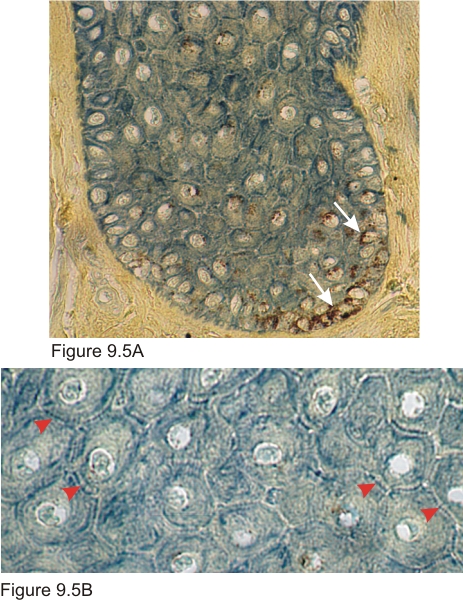
Figure 9.5A Section of thick skin stained with TPA.

This method selectively stains the keratin filaments green. These filaments are abundant in the cytoplasm of keratinocytes, which explains the deep staining of the epidermis in this section.

Some basal cells contain melanin granules (arrows).

Figure 9.5B Higher magnification of a thin section of thick skin stained with TPA as in Figure 9.5A.

The keratin filaments are inserted on the desmosomes of keratinocytes and form spindles or spines (arrowheads) at the interfaces.

These spines seen at the interfaces of polyhedral keratinocytes are responsible for the term stratum spinosum given to this layer of the epidermis.

Stain: TPA
Magnification: ×900 (Fig. 9.5A); ×1200 (Fig. 9.5B)


We'd very much appreciate receiving your comments and suggestions!We'd very much appreciate receiving your comments and suggestions!

Creative Commons Licence The text and images of this Histology Atlas, by Yves Clermont, Michael Lalli & Zsuzsanna Bencsath-Makkai, are licensed under a Creative Commons Attribution-Noncommercial-No Derivative Works 2.5 Canada Licence and cannot be modified without the written permission of the authors. Use of any text or images must carry an acknowledgement which includes a link to the original work.


HAweb/Zs B-M -> HAbase-generated on 2013-09-13