Contents Yves Clermont, Michael Lalli, Zsuzsanna Bencsath-Makkai
LIGHT-MICROSCOPIC HISTOLOGY ATLAS
     18. Eye
  1     2     3     4     5     6     7     8     9     10     11     12     13     14     15     16     17     18     19     20     21     22     23     24     25  
  26     27     28     29  

    18.29   Retina and optic nerve Version française
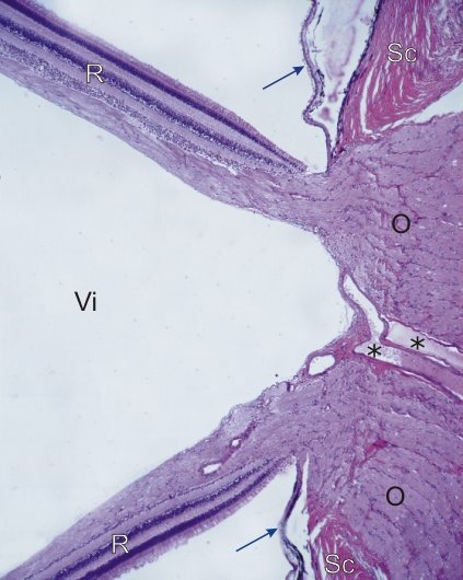
Section of the optic nerve at the back of the eye cavity of a dog. This field corresponds to the framed area D in Figure 18.1.

This field shows the retina (R), detached here artificially from the choroid (arrows).

The retina is continuous with the optic nerve (O) at the exit of the ocular globe. Large vessels (*) are seen in the centre of the optic nerve.

The sclera (Sc) and the space occupied by the vitreous body (Vi) are labelled.

Stain: H–­­E
Magnification: ×70


We'd very much appreciate receiving your comments and suggestions!We'd very much appreciate receiving your comments and suggestions!

Creative Commons Licence The text and images of this Histology Atlas, by Yves Clermont, Michael Lalli & Zsuzsanna Bencsath-Makkai, are licensed under a Creative Commons Attribution-Noncommercial-No Derivative Works 2.5 Canada Licence and cannot be modified without the written permission of the authors. Use of any text or images must carry an acknowledgement which includes a link to the original work.


HAweb/Zs B-M -> HAbase-generated on 2013-09-13