|
||
| 18. Eye | ||
| 1 2 3 4 5 6 7 8 9 10 11 12 13 14 15 16 17 18 19 20 21 22 23 24 25 | ||
| 26 27 28 29 |
| |||
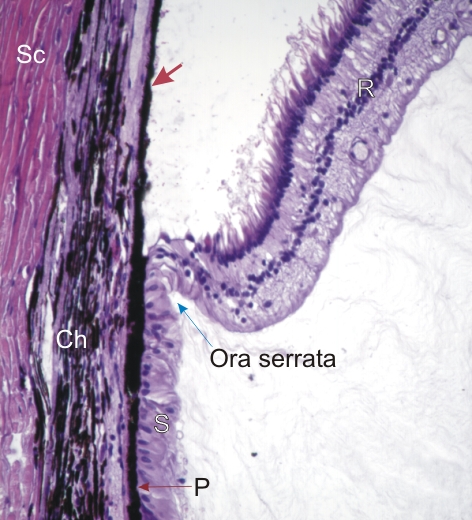 |
This field shows the borderline, called ora serrata, between the ciliary body and the retina of a dog (see Figure 18.10).
The lining of the ciliary body shows its basal pigmented cell layer (P) and the non-pigmented cell layer at its surface (S). The pigmented cell layer of the ciliary body is continuous with the pigmented cell layer of the retina (arrow). The non-pigmented cell layer of the ciliary body is continuous with the neural retina or retina proper (R). In this section the retina is artificially separated from the pigmented cell layer of the retina. The choroid (Ch) and sclera (Sc) are also identified. Stain: HE
|
||