Contents Yves Clermont, Michael Lalli, Zsuzsanna Bencsath-Makkai
LIGHT-MICROSCOPIC HISTOLOGY ATLAS
     18. Eye
  1     2     3     4     5     6     7     8     9     10     11     12     13     14     15     16     17     18     19     20     21     22     23     24     25  
  26     27     28     29  

    18.18   Lense and a ciliairy process Version française
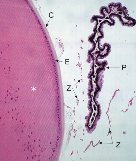
Section of a lens and of a ciliary process.

The lens shows a thick capsule (C), the anterior epithelium of the lens (E) and closely packed lens fibres (*). The posterior surface of the lens is not covered with such an epithelium. The lens, on the other hand, is completely covered by the capsule.

The oblique section of the ciliary process (P) shows an epithelium composed of two layers of cells at the surface of a thin layer of connective tissue.

The basal cells of this epithelium are heavily pigmented, while the surface cells are not (see the following figures).

A few zonular fibres (Z) are identified.

Stain: H–­­E
Magnification: ×250


We'd very much appreciate receiving your comments and suggestions!We'd very much appreciate receiving your comments and suggestions!

Creative Commons Licence The text and images of this Histology Atlas, by Yves Clermont, Michael Lalli & Zsuzsanna Bencsath-Makkai, are licensed under a Creative Commons Attribution-Noncommercial-No Derivative Works 2.5 Canada Licence and cannot be modified without the written permission of the authors. Use of any text or images must carry an acknowledgement which includes a link to the original work.


HAweb/Zs B-M -> HAbase-generated on 2013-09-13