|
||
| 18. il | ||
| 1 2 3 4 5 6 7 8 9 10 11 12 13 14 15 16 17 18 19 20 21 22 23 24 25 | ||
| 26 27 28 29 |
| |||
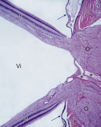 |
Coupe du nerf optique au fond de lœil dun chien. Ce champ correspond à lencadré D de la figure 18.1. Ce champ montre la rétine (R), détachée artificiellement de la choroïde (flèches). La rétine est continue avec le nerf optique (O) à sa sortie du globe occulaire. De gros vaisseaux (*) sont présents dans le centre du nerf optique. La sclérotique (Sc) et lespace occupé par le corps vitré (Vi) sont identifiés. Coloration: HÉ
|
||