|
||
| 18. Eye | ||
| 1 2 3 4 5 6 7 8 9 10 11 12 13 14 15 16 17 18 19 20 21 22 23 24 25 | ||
| 26 27 28 29 |
| |||
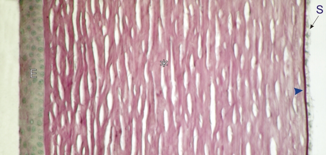 |
Cornea of the eye of a dog.
At the surface of the cornea the thin epithelium (E) is stratified squamous. Underneath the epithelium the thick layer of dense connective tissue (*) is formed of type I collagen fibres and fibrocytes embedded in an amorphous ground substance rich in proteoglycans. These glycoproteins are partly responsible for the PAS staining of this connective tissue. This ground substance may be responsible for the transparency of the cornea. On the inner aspect of the cornea and facing the anterior chamber of the eye, a simple epithelium (S) sits on the thick Descemets membrane (arrowhead). This membrane, which is equivalent to a thick basement membrane, is intensely stained purple with PAS. Stain: PAS-Hematoxylin
|
||