|
||
| 18. Eye | ||
| 1 2 3 4 5 6 7 8 9 10 11 12 13 14 15 16 17 18 19 20 21 22 23 24 25 | ||
| 26 27 28 29 |
| |||
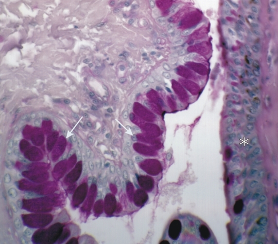 |
Conjunctiva of a dogs eye stained with PAS.
(Left) This field shows the palpebral conjunctiva with its epithelium containing numerous PAS-positive goblet cells (arrows). (Right) The bulbar conjunctiva (*) shows a stratified epithelium with only a few epithelial mucous cells. The image emphasizes the fact that the composition of the epithelium along the conjunctiva is highly variable. Stain: PAS-Hematoxylin
|
||