Contents Yves Clermont, Michael Lalli, Zsuzsanna Bencsath-Makkai
LIGHT-MICROSCOPIC HISTOLOGY ATLAS
     18. Eye
  1     2     3     4     5     6     7     8     9     10     11     12     13     14     15     16     17     18     19     20     21     22     23     24     25  
  26     27     28     29  

    18.5   Eyelid Version française
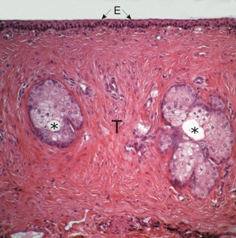
Section of an eyelid.

The plane of cut is horizontal through this eyelid. This field shows two transversely cut sebaceous Meibomian glands. Their central canals (*) are indicated.

These glands are located in the dense connective tissue of a tarsal plate (T). These plates give rigidity to the eyelids and serve as the insertion sites of muscles that permit the opening and closing of the eye.

The epithelium (E) of the palpebral conjunctiva is also identified.

Stain: H–­­E
Magnification: ×25


We'd very much appreciate receiving your comments and suggestions!We'd very much appreciate receiving your comments and suggestions!

Creative Commons Licence The text and images of this Histology Atlas, by Yves Clermont, Michael Lalli & Zsuzsanna Bencsath-Makkai, are licensed under a Creative Commons Attribution-Noncommercial-No Derivative Works 2.5 Canada Licence and cannot be modified without the written permission of the authors. Use of any text or images must carry an acknowledgement which includes a link to the original work.


HAweb/Zs B-M -> HAbase-generated on 2013-09-13